Lumps beneath the skin are a common concern and in many cases, they turn out to be benign conditions such as lipomas. While these fatty growths are usually harmless, changes in size, appearance or sensation can understandably cause worry. Because lipomas tend to grow slowly and are often painless, they are sometimes left unattended for years. However, timely assessment is important, not only to confirm the diagnosis, but also to determine whether removal may be beneficial for comfort, function or cosmetic reasons.
This case study highlights Dr Aaron Poh’s patients' experience underscores the value of early evaluation and how a straightforward procedure can provide reassurance and a good clinical outcome.
The patient was a 42-year-old woman who presented with a lump over her left forearm. She had first noticed the swelling some time ago, but it had gradually increased in size.
While the lump was not painful, it had started to:
On further discussion, there was no history of trauma or infection in the area. However, the steady growth of the lump made it important to assess it properly.
This presentation is typical of many patients with lipomas, where the absence of pain can delay consultation, even as the lump continues to enlarge over time.
On examination, the lump appeared:
These characteristics are typical of a lipoma, a benign growth made up of fatty tissue. Lipomas are generally harmless and slow-growing, but removal may be recommended if they increase in size, cause discomfort or affect appearance.
After a thorough assessment and discussion, the provisional diagnosis of a lipoma was made.

The patient was counselled on management options, including observation versus surgical removal. Given the increase in size and her concerns, she opted for surgical excision.
The procedure was planned with the following goals in mind:
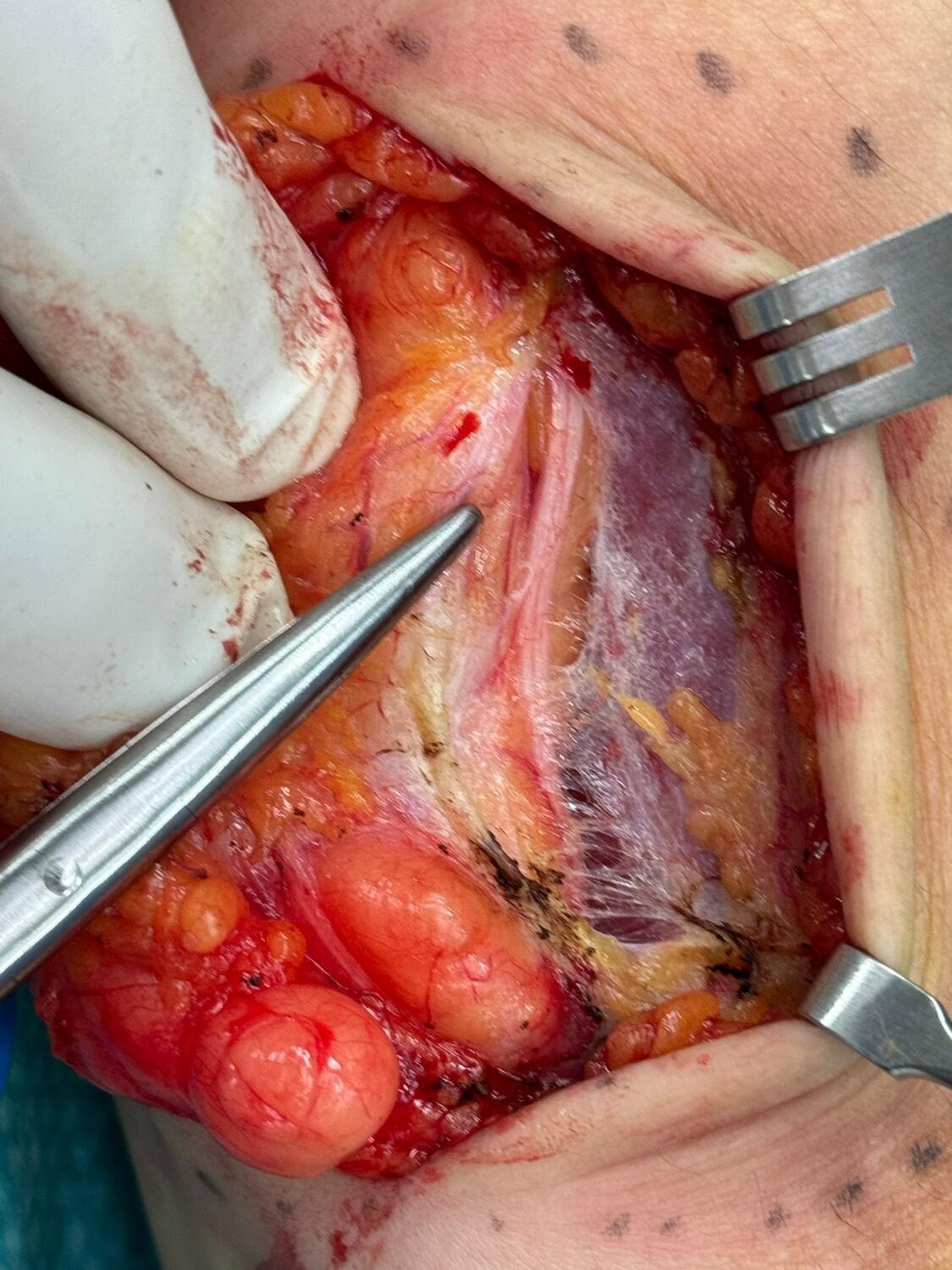
The excision was performed in a controlled clinical setting under appropriate anaesthesia to ensure comfort throughout the procedure.
A small incision was made directly over the lump, allowing careful access to the underlying tissue. The lipoma was gently separated from surrounding structures and removed in its entirety. The wound was then meticulously closed to support optimal healing and minimise scarring.
The excised tissue was sent for histological examination as a routine precaution.
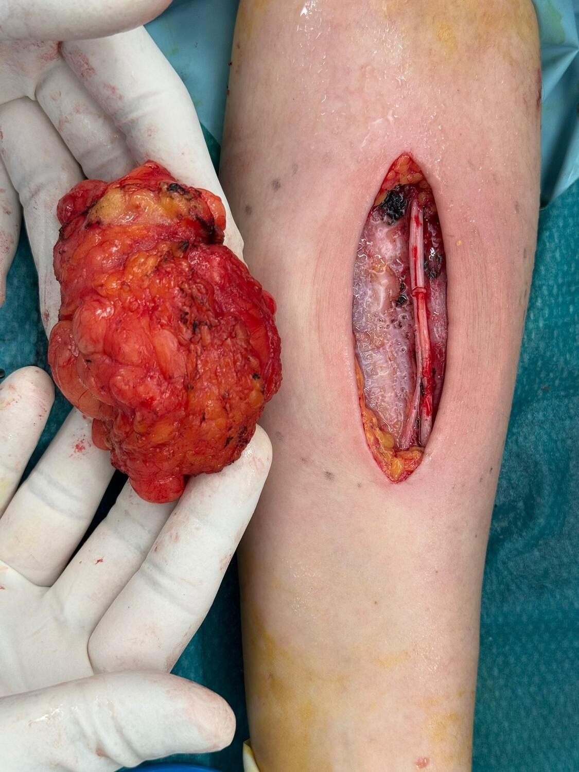
During the procedure, the mass was found to be:
There were no features to suggest invasion or malignancy.
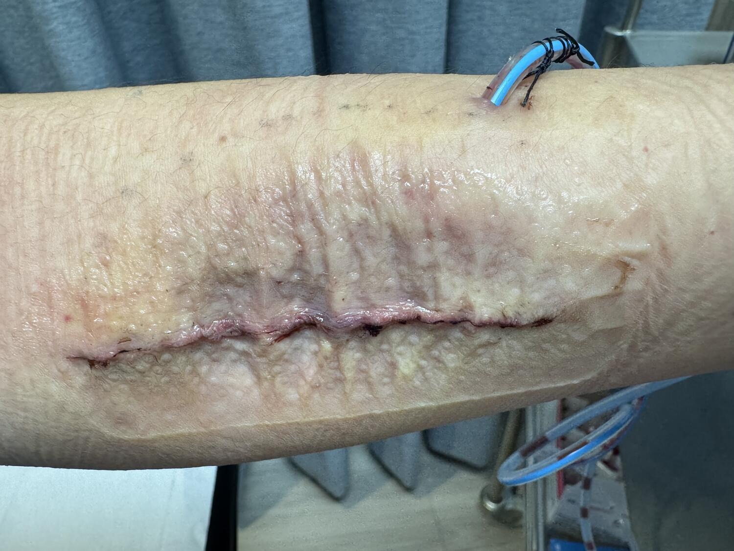
The patient’s recovery was smooth and uneventful.
In the immediate post-operative period, she experienced only mild discomfort, which resolved quickly. The wound healed well, with no signs of infection or complications.
At her 2.5-month follow-up, the outcome was very encouraging:
The contour of the forearm appeared natural and improved
The surgical site had healed neatly
There was no evidence of recurrence

The procedure achieved a successful outcome, both medically and aesthetically. The lipoma was completely removed, symptoms were resolved and the patient was pleased with the result.
If you or a loved one has been suffering from lumps, we encourage you to schedule an appointment to have the lump assessed and treated.
Patient details have been anonymised and modified to protect confidentiality. The information presented is for general educational purposes.


Copyright © Alpine Surgical Practice | Terms & Conditions


